A direct comparison of open and robotic-assisted surgery
Let’s look at what a conventional radical prostatectomy and a robotic-assisted radical prostatectomy involve, and compare the two methods.
In the open surgical method, the surgeon accesses the prostate by making an 8 to 10 cm incision in the lower abdomen, above the pubic bone. In robotic-assisted surgery, the patient is placed in the lithotomy position, with their legs raised above their torso The prostate is accessed via six small incisions in the abdomen. The first small incision, just above the navel, allows the surgical team to fill the patient’s abdomen with carbon dioxide to improve their field of vision. A trocar (insertion aid) is then used to insert the camera through the incision, with further trocars placed in other incisions.
- Open: One incision in the lower abdomen, 8 to 10 cm in length
- Robotic-assisted: 6 small incisions
- Open: Standard position, lying horizontally
- Robotic-assisted: Lithotomy position, patient’s legs raised above their torso, abdomen filled with gas
The first phase of the operation involves carefully exposing the prostate. The two surgical methods differ in terms of the sequence of steps that follow. The open surgical method starts by detaching the prostate, which is embedded in the pelvic floor and the pelvic muscles. The robotic-assisted surgical method accesses the prostate from a different direction. The surgeon starts by detaching the urethra directly above the prostate, releasing the seminal vesicles and severing the seminal duct. It then detaches the prostate from the pelvic floor, pelvic muscles and the rectum.
The next step – if deemed reasonable from an oncological perspective – is nerve sparing. This time-consuming step is performed with great caution. Firstly, the powerful veins need to be ligated. The layer containing vascular and nerve tissue is separated from the prostate millimetre by millimetre. Special attention is paid to ensuring that no electrical energy or heat is applied in this step, as this could damage the sensitive nerves.
- Open: Surgical loupes with 4x to 5x magnification
- Robotic-assisted: 3D camera with 10x magnification
After completing the nerve-sparing step, the prostate is completely separated from the urethra. In doing so, the urethral sphincter is fully preserved using a technique developed by doctors at the Martini-Klinik in order to preserve the patient’s continence.
- Open: Blood loss 550–750 mL, transfusion rate 3.5%
- Robotic-assisted: Blood loss 150–250 mL, transfusion rate 1.5%
It is only medically possible to spare the nerves if it can be ascertained that the tumour has not extended beyond the capsule. It is not possible to determine this in the operating theatre without a microscope. This is why, during each prostatectomy at the Martini-Klinik, the removed prostate is sent to the pathology department and histologically examined using the NeuroSAFE technique – a technique that we developed and published. The bundle of nerve fibres is only removed if cancer cells are detected where the nerve fibres connect to the prostate.
Special surgical techniques used at the Martini-Klinik to preserve potency and continence
The urinary tract is then reconnected by suturing the urethra and the bladder neck together and then splinted with an indwelling catheter. The suture affects as little of the sphincter tissue as possible in order to preserve this muscle, which is responsible for continence.
In most but not all cases, a drainage tube is also inserted to remove exudate and lymphatic fluids. The abdominal incision, or the several small abdominal incisions, are then closed.
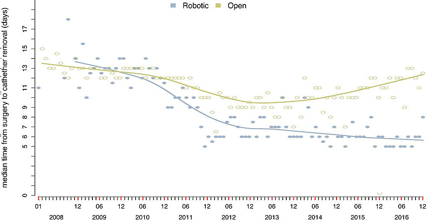
- Open: Catheter remains in place for 12 days on average
- Robotic-assisted: Catheter remains in place for 7 days on average
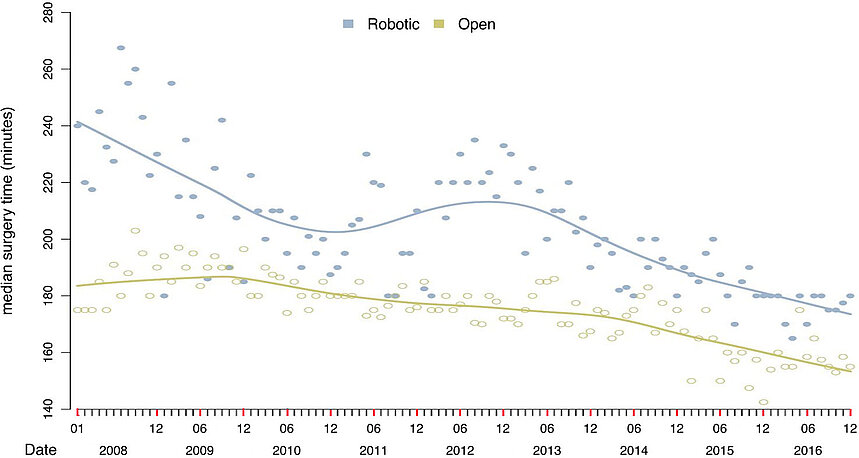
Nowadays, there is no significant difference in the duration of the two surgical methods.
- Open: Surgery takes 175 minutes on average
- Robotic-assisted: Surgery takes 195 minutes on average
The final step in the surgery is to close the wounds.