We offer this method to help you and your urologist to gain clarity where initial findings have been inconclusive.

MRI fusion biopsy with image recording
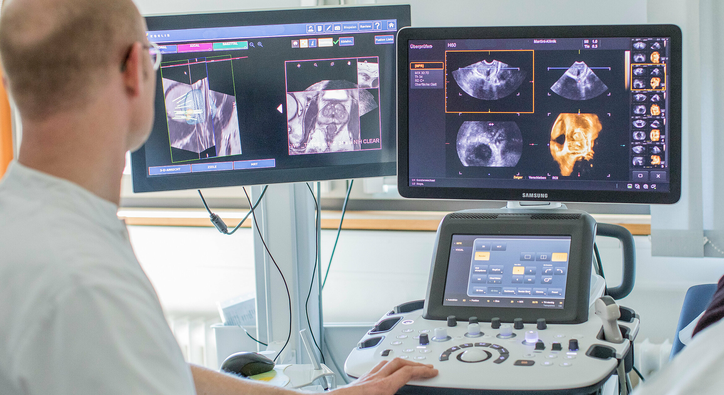
In a fusion biopsy, a conventional ultrasound image is used to create a three-dimensional image of the prostate in a matter of seconds. This 3D image is then fed into the live image (3D TRUS biopsy). At the same time, the image from the magnetic resonance imaging (MRI) scan is combined with the live ultrasound image, with both images precisely laid over each other. A fusion biopsy is therefore better able to visualise certain anatomical and functional aspects of defined areas within the prostate. Information about suspect areas generated by both techniques can then be used in combination during the biopsy.
A fusion biopsy makes it easy to detect altered tissue, which enables the doctor to take samples using the biopsy needle with greater precision. Making digital records of the biopsy removal sites also makes it possible to revisit the locations of abnormal findings, such as during a re-biopsy in an active surveillance schedule.
At the Martini-Klinik, we can perform MRI-guided fusion biopsy using a range of instruments as well as transrectal biopsy via the rectum and transperineal biopsy via the perineum.
To help plan a fusion biopsy, we send the MRI images to the Radiology department at UKE for analysis and assessment. This takes a few days. If you have your MRI scan at UKE, the Radiology department can analyse the images and send us an electronic patient record without delay.
Has MRI-guided fusion therapy improved diagnostic processes?
Fusion biopsy is a way to provide clarity at an early stage, especially if there is reason to suspect atypical localisation of a tumour, and facilitate precise repeat biopsies in active surveillance schedules. Detecting higher-grade tumours, also known as significant tumours, is particularly important.
Fusion imaging makes it possible to call up the 3D fusion image before a re-biopsy or in the event that an MRI scan returns new findings, thereby enhancing the precision of re-biopsies.
Studies show better detection of clinically significant tumours
A prominent study (known as the PRECISION trial) published in the New England Journal of Medicine compared standard biopsies with fusion biopsies. The results showed that the clinically significant tumours were detected more effectively by the fusion biopsy method (38%) compared to standard biopsies (26%). Following confirmation of these findings by various working groups, MRI-guided fusion biopsy has now become an established and vital method in the diagnosis of prostate carcinoma.
Your urologist will discuss with you how to approach the findings, which often do not require immediate treatment, taking into account various factors such as your age, any secondary illnesses and your family’s history of cancer.
Far fewer non-significant tumours identified by chance
The phenomenon in which MRI-guided fusion biopsies detect significant tumours more frequently and non-significant tumours less frequently is backed up by our own experience of over 4,000 MRI-guided fusion biopsies carried out at the Martini-Klinik. This includes many tumours localised in the prostate that would have been difficult or near-impossible to detect in an ultrasound biopsy due to calcification in the prostate or the size of the prostate.
Prof. Dr. Lars Budäus
Summary
In diagnostically complicated cases, such as when patients have a family history of prostate cancer, or if imaging techniques have returned unclear or contradictory findings, the close cooperation we enjoy with our colleagues in the Radiology department, the Nuclear Medicine department and the Institute of Human Genetics can often deliver an individual diagnosis.
Diagnostic consultation
+49 (0)40 7410-28672
+49 (0)40 7410-40245
mk-diagnostik@uke.de
In a fusion biopsy, a conventional ultrasound image is used to create a three-dimensional image of the prostate, which is fed into the live image (3D TRUS biopsy). At the same time, the MRI image is combined with the live ultrasound image, with both images precisely laid over each other. The information from both techniques can be used in combination during the biopsy to gain a better understanding of changed tissue.
A fusion biopsy is a way to achieve clarity at an early stage and conduct precise re-biopsies in the case of active surveillance. Studies have shown that fusion biopsy is better at identifying clinically significant tumours.
