A subsidiary of the University Medical Center Hamburg-Eppendorf

Ein Arzt zeigt auf den Monitor eines Ultraschallgeräts

The path to a reliable diagnosis

Prostate diseases become more likely as a person gets older. Screening is recommended in men aged 45 and over. The earlier a tumour is identified, the better the chances of curing it. If the tumour is growing within the prostate (i.e. confined to the prostate), the chances of recovery are well over 95% when treated effectively. However, few men in the at-risk group actually undergo annual cancer screening. And, despite intensive research in this field, there are still no clear markers that allow us to detect prostate carcinoma.

Screening

Screening for prostate cancer usually involves palpation (physical examination) and an ultrasound. In a transrectal ultrasound (TRUS), the transducer is inserted via the back passage (rectum). We also recommend arranging a blood test with your urologist or GP to determine your PSA level. Palpation allows your doctor to identify changes to the prostate. Your PSA level provides an indication about the level of prostate-specific antigens in your blood. If this is elevated, it could indicate a prostate carcinoma – however, it is important to remember that other factors can also lead to an elevated PSA level.

If your PSA level is abnormally high or your urologist identifies abnormal stiffness when examining your prostate, this provides reason to suspect a prostate carcinoma. In this case, the next step is to take a sample of the prostate tissue (biopsy) and examine it. This allows your doctor to determine whether a malignant tumour is present or not.

Martini-Klinik

Accompanies you through this topic

Prof. Dr. Georg Salomon
Faculty member

A biopsy is the next step towards diagnosis

If there is reason to suspect you have a prostate carcinoma (due to an elevated PSA level and/or findings from palpation of the prostate), the next step is usually taking a sample of tissue from the prostate. Due to the prostate’s location in the body, different methods are used to access it.

Transrectal prostate biopsy 

Biopsies of the prostate are currently often conducted via the back passage (rectum) under local anaesthetic in what is known as a transrectal ultrasound scan (TRUS) biopsy. In order to reduce the risk of infection, it is recommended practice to test for antibiotic resistance in the intestinal bacteria so that antibiotic treatments can be adjusted if necessary. An alternative procedure is perineal biopsy, also performed under local anaesthetic.

Perineal prostate biopsy

In this procedure, the biopsy is taken via the perineum under local anaesthetic. This method carries an exceptionally low risk of infection, making it an ideal choice if a patient has specific infection risk factors. Such factors include resistance against common antibiotics being identified in a rectal swab, diabetes mellitus, patients who take immunosuppressives, prostate inflammation and past surgeries on the rectum. The risk of infection is lower in perineal biopsies because the intestinal mucosa is not perforated, so there is less chance of intestinal bacteria spreading into the prostate. 
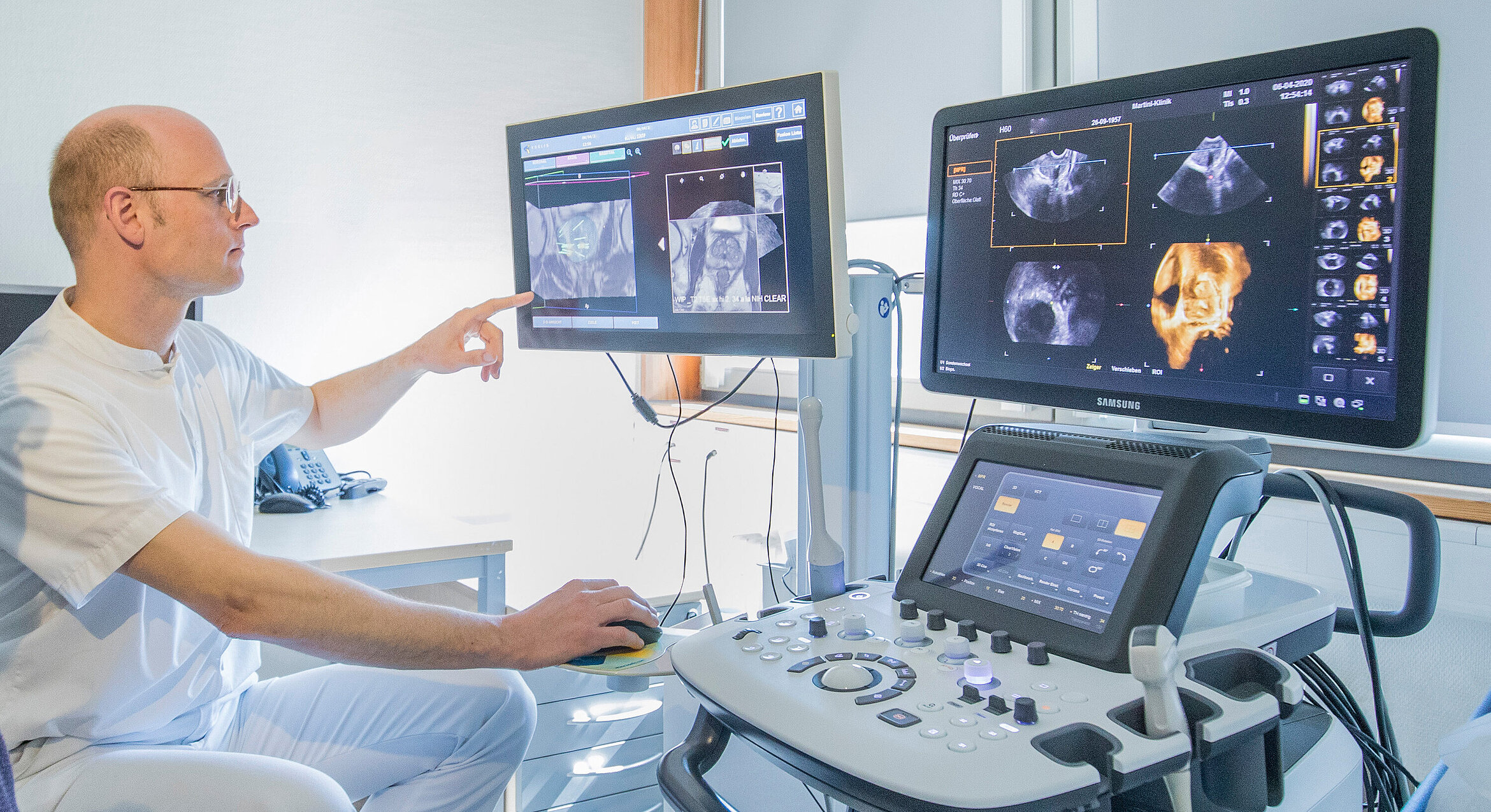
Fusion biopsy

After an ultrasound-based biopsy, if no tumour has been identified but a patient’s PSA level is still significantly elevated, German guidelines currently recommend performing an MRI scan before a further prostate biopsy.  This procedure helps to examine areas of the prostate where cancer is suspected and take targeted samples if suspected tumour tissue is identified in the prostate.

Based on European guidelines and the added diagnostic value of such scans, prostate MRIs are now commonly performed before the initial biopsy. By visualising areas of the prostate where cancer is suspected, MRI scans make it possible to achieve a higher detection rate and assess risks more effectively.

Find out more about fusion biopsies

It’s cancer. What’s next?

If you are diagnosed with prostate cancer, the next step is to conduct further tests to assess how aggressive your tumour is and how far it has spread. This information is essential to determine the best possible treatment options in your case. In the case of a slow-growing tumour that has not yet metastasised, active surveillance might be the best choice.

Firstly, a tissue sample enables us to determine a Gleason score and TMN staging. Bone scintigraphy allows us to determine whether the cancer has already spread to the bone system, which is the most common form of metastatic spread for prostate carcinoma.

Once all the tests and scans have been completed, the doctor and patient discuss suitable treatment options.

How is prostate cancer diagnosed?

In addition to palpation and a transrectal ultrasound (TRUS), it is also important to determine the patient’s PSA level. 
If these examinations return abnormal findings and prostate cancer is suspected, further diagnostics should be conducted, namely a biopsy. There are different types of biopsy.

Transrectal or transperineal prostate biopsy

In a transrectal or transperineal prostate biopsy, a special hollow needle is used to take small samples of tissue from the prostate, which are then checked for cancer cells. 

Fusion biopsy

A fusion biopsy involves an MRI scan prior to the biopsy. This provides a view of areas of the prostate where cancer is suspected. In a fusion biopsy, the MRI image is fused with the ultrasound image during the biopsy to increase the detection rate.